We all have seen a microscope before. Maybe a very simple one at home when we were kids to look at leaves or onion skin, at school (if we were lucky!), or on the FBI programmes on TV! But, how did microscopy start and how does a microscope work?
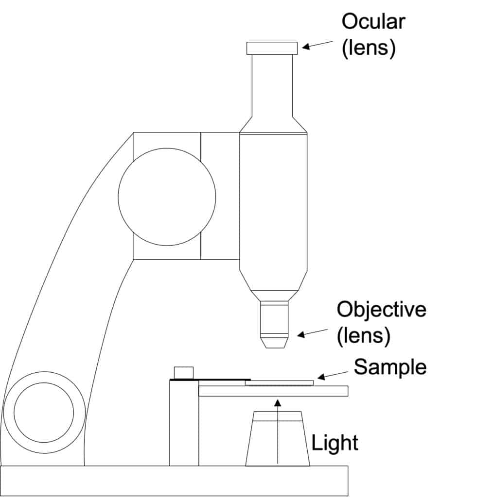
Probably, when we think about microscopy, the first thing that comes to mind is a light microscope. In this kind of microscopy, the sample is hit by light (photons), which will then be reflected and pass through the microscope lenses which will magnify the image that is too small to be seen with the naked eye.
Nevertheless, there are several types of microscopes that currently exist, and not all of them use light! So, if we had to choose only one word to generally define a microscope, we would probably pick ‘lens’. Lenses were produced hundreds of years before microscopes existed, and there is evidence that small magnifying lenses had been used even in Roman times, mainly for refine artwork. It was not until the 13th century, however, that lenses were used for other purposes, such as to correct eye vision. In 1590, two spectacle makers, Zaccharias Janssen and Hans Lipperhey, are thought to be the first people to have developed the concept of compound microscope (i.e., which uses more than one lens). Nevertheless, it was not until 1625 that the word ‘microscope’ was first used to described Galileo’s compound microscope, created in 1609 [1].
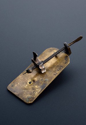
Despite the existence of these instruments, there are no recollections of observations made with them until later that century. Two contemporary people, Antonj van Leeuwenhoek (Dutch) and Robert Hook (English), were the first people to create a collection of detailed illustrations using microscopes. Leeuwenhoek was a draper, and he used lenses to look at the threads in cloth. He created the impressive amount of 500 different single lens microscopes and started to use them to look at different things, like a drop of water. By looking into pond water, he discovered microorganisms, which earned him the title of the father of microbiology [2].
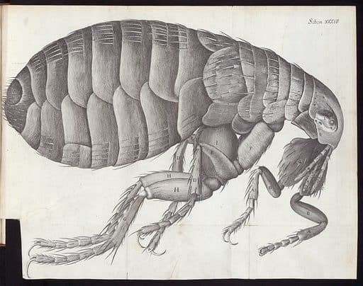
At the same time in England, Robert Hooke used a compound microscope to study details from organisms and plants that were completely unknown before. He published all his work in 1665 in a book called ‘Micrographia’ [3]. He also invented pieces that would become components of modern light microscopes, such as the iris diaphragm. The work of both Leeuwenhoek and Hook was essential for the establishment of cell theory later on.
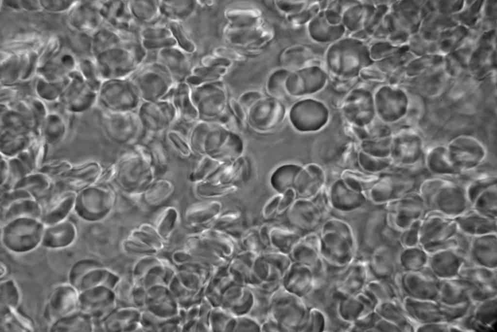
In 1934, another Dutch, Frits Zernike, described for the very first time the concept of phase contrast microscopy [4]. Contrast is defined by the signal (colour or brightness) differences between the background and the tissue of interest. Zernike realised that it was possible to use this property to generate images from unstained, colourless samples.
Together with the improvements on microscopy, the appearance of microtomes to cut thinner sections, as well as staining methods, started making it possible to differentiate structures in the tissues. One of the first documented stainings was done by Joseph von Gerlach, who managed to differentiate for the first time the nucleus from the cytoplasm of cells by using carmine (a natural pigment obtained from small insects). Another well-known staining example is the Gram staining (1884), developed by Hans C. Gram, which can be used to differentiate classes of bacteria.
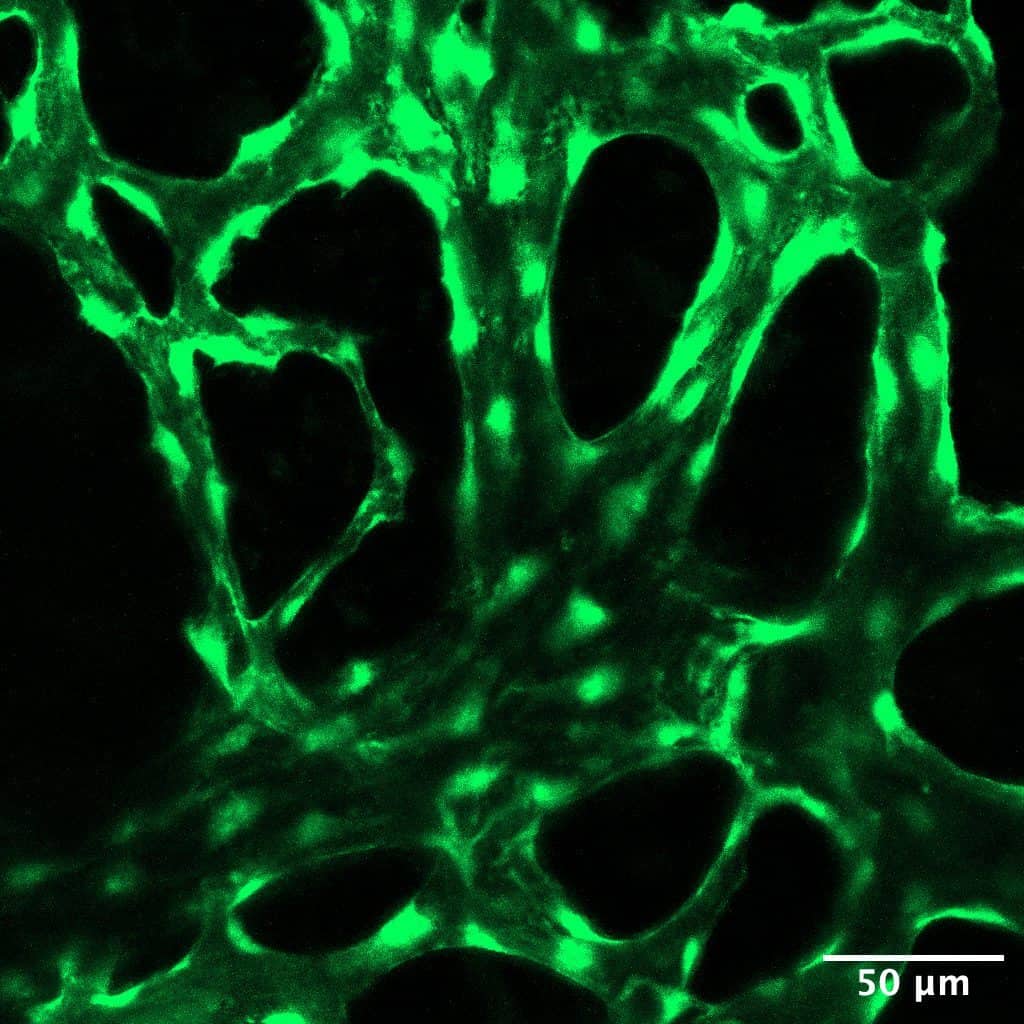
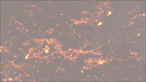
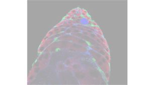
Many of these early staining techniques are still widely use in the labs [5], but it was the utilisation of fluorescence what revolutionised light microscopy. Fluorescent proteins are excited and emit at different wavelengths. In 1871, the first fluorescent protein, fluorescein, was used and, since then, many others have been identified and isolated. Some of the most commonly used fluorescent proteins are the green fluorescent protein (GFP) or dsRed, which were first isolated from jellyfish and coral, respectively. Nowadays, it is possible to couple the expression of these fluorescent proteins to either antibodies for identification of specific cell populations or to specific genes for the study of expression levels. Currently, there are different fluorescent microscopes such as epi-fluorescent, confocal, or two-photon microscopes [6].
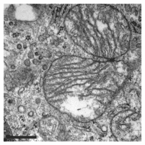
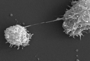
In 1931, Max Knoll and Ernst Ruska overcame the resolution and magnification limits of light by creating the first Transmission Electron Microscope (TEM). In this case, a beam of electrons, instead of photons, is used to image the samples. As the wavelength of electrons is shorter than the light, the acquisition of very detailed magnified images is achieved. In 1965, the first Scanning Electron Microscope (SEM) was developed, which allowed for the acquisition of 3D images of surfaces in comparison to the 2D images of the sample that are created with TEM [7].
In 1981, a new phase for microscopy started with the invention of the Scanning Tunnelling Microscopy (STM), which allows for imaging at an atomic level. This new microscopy technique was developed by Gerd Binning and Heinrich Rohrer. That same year, Binning, together with Calvin Quate and Christoph Gerber, developed the Atomic Force Microscopy (AFM). These two scanning probe microscopy techniques, which allow for the observation of individual atoms, were tightly knitted to the development of nanotechnology [8].
Microscopy has become a very important tool to better understand the world around us: from learning how our body works and how diseases affect us to the study and development of new materials, among many other applications. As microscopy keeps evolving, we cannot wait to see what other new features will be implemented to show us the unseen!
By Sara González Antón, PhD student at Imperial College London.
More information:
- Wollman et al., 2013.
- Lane 2015.
- Micrographia, by Robert Hooke, 1665. Available online here.
- Introduction to Phase Constracts Microscopy. Source: Microscopyu. Available online here.
- An Introduction to Routine and Special Staining. Source: Leica Biosystems. Available online here.
- Microscopy Techniques. Source: Microscopyu. Available online here.
- Transmission Electron Microscopy vs Scanning Electron Microscopy. Source: ThermoFisher. Available online here.
- Baird and Shew, 2004.
- The microscope. Source: Science Museum. Available online here.