The secluded Baobab
– Luis Moliner Cachazo, King’s College London.
Winner of Technique and quality of the photograph category

When doing fieldwork in the Okavango Delta (Botswana) and despite my research being on aquatic insects, I really wanted to spot the most emblematic tree in Africa, the Baobab (Adansonia digitata). However, these trees are not very common in this area, so my collaborators stopped the boat for me to capture this majestic specimen with a little bird on top.
Uncharted Planet
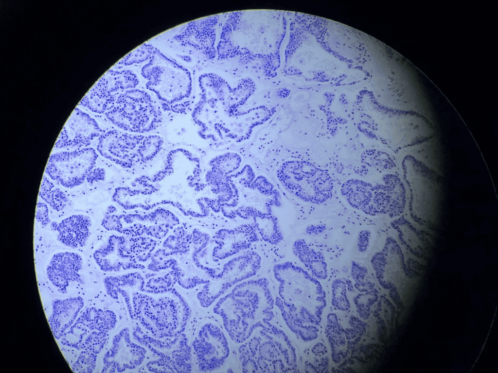
– Adrian Acuña, Memorial Sloan Kettering Cancer Center
Winner of Originality and artistic character of the image
Photograph of a mouse thyroid carcinoma stained with hematoxylin and eosin. Histology is fundamental in medical diagnosis since it allows visualizing the structure of the tissue and the characteristic changes that the tissue may have undergone. In addition to this, the use of genetically engineered mouse models provides an opportunity to explore the unknown biology behind cancer.
Programming immune cells to kill cancer
– Luisa Chocarro de Erauso, Navarrabiomed-UPNA-IdiSNA
Winner elected by SRUK/CERU members
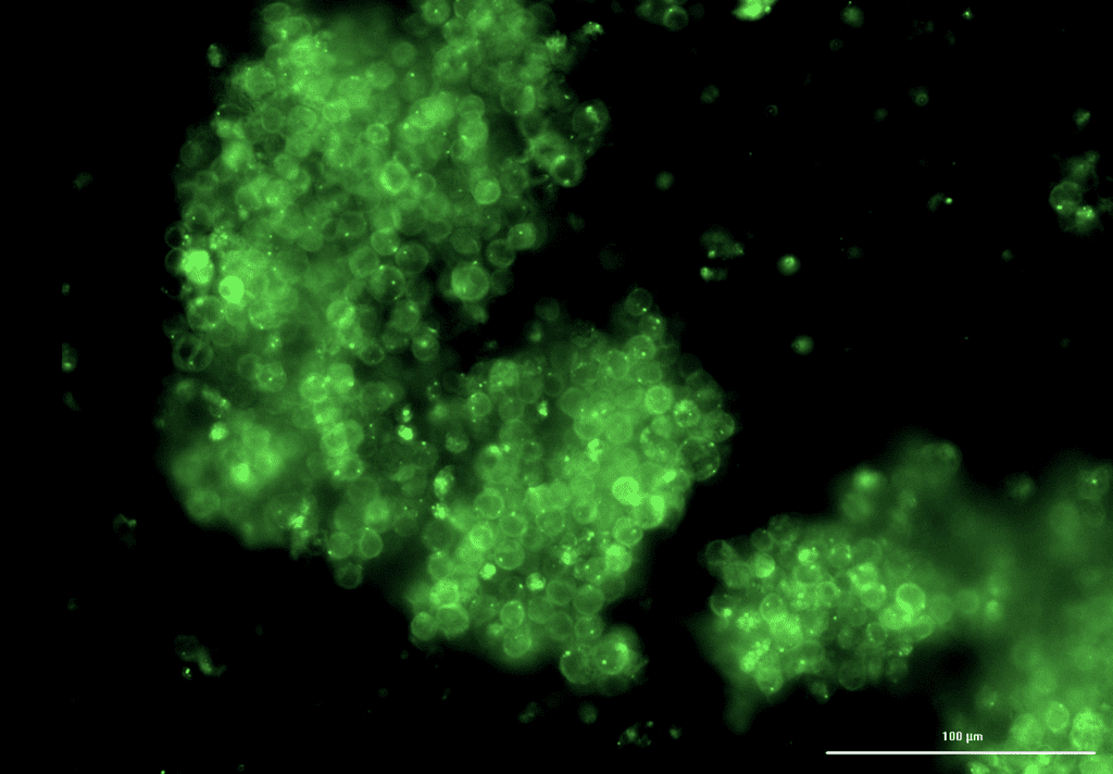
Can we programme immune cells to kill cancer? This microscopy image proves it is possible. Here we can see T-cells, immune system soldiers, genetically modified to express an activating molecule fused to a green fluorescence molecule. It represents the possibility to engineer CAR T-cells, cancer-fighting immune super soldiers, and one of the most promising advanced therapies for personalised cancer treatment.
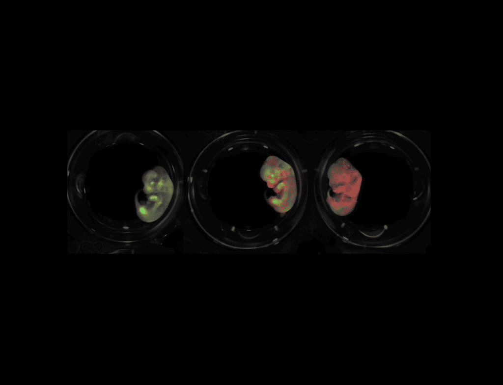
Colouring ontogeny
– Beatriz Fernandez Ruiz, Imperial College London.
Winner of Research Communication
Bioluminescence imaging of dystrophin, in green, and utrophin, in red, in single- (left and right panels) and dual-reporter (middle panel) E13 mouse embryos. Dystrophin and utrophin are key proteins for Duchenne Muscular Dystrophy (DMD), which is a lethal muscle wasting disorder affecting 1 in 3000 boys. In vivo bioluminescence imaging offers a platform to study simultaneously the expression patterns of DMD’s key proteins during development in order to uncover novel therapeutic targets.
Targeting Nanomaterials
– Sara Escalera, University College London
EpCAM targeting nanomaterials interacting with the surface of patient derived pancreatic cancer cells (PDX185), which overexpress this cancer-related marker. Red: phalloidin, blue: DAPI stained nuclei, green: targeted nanomaterials.
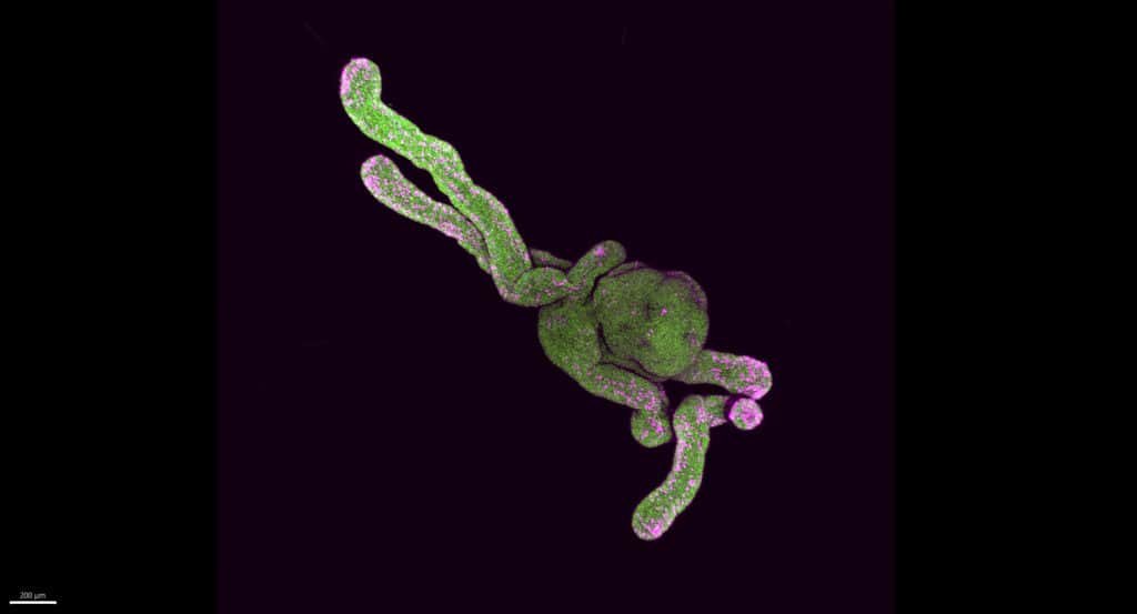
Octopus organoid
– Carmen Moreno Gonzalez, The Francis Crick Institute
3D render of a neural crest organoid reminiscent of an octopus. Cell nuclei are labelled in green and neural crest cells in pink. Neural crest organoids are derived from pluripotent human cells and mimic processes which happen during early embryonic development. Organoids are particularly powerful tools for disease research and drug screening.
Immunofluorescence of human glioblastoma cells
– Maria Rosado Rodriguez, University College London
Confocal microscopy image of an immunofluorescence co-distribution between two proteins, WIP in green and IQGAP in blue together with actin filaments in red, in human glioblastoma cells (U87).
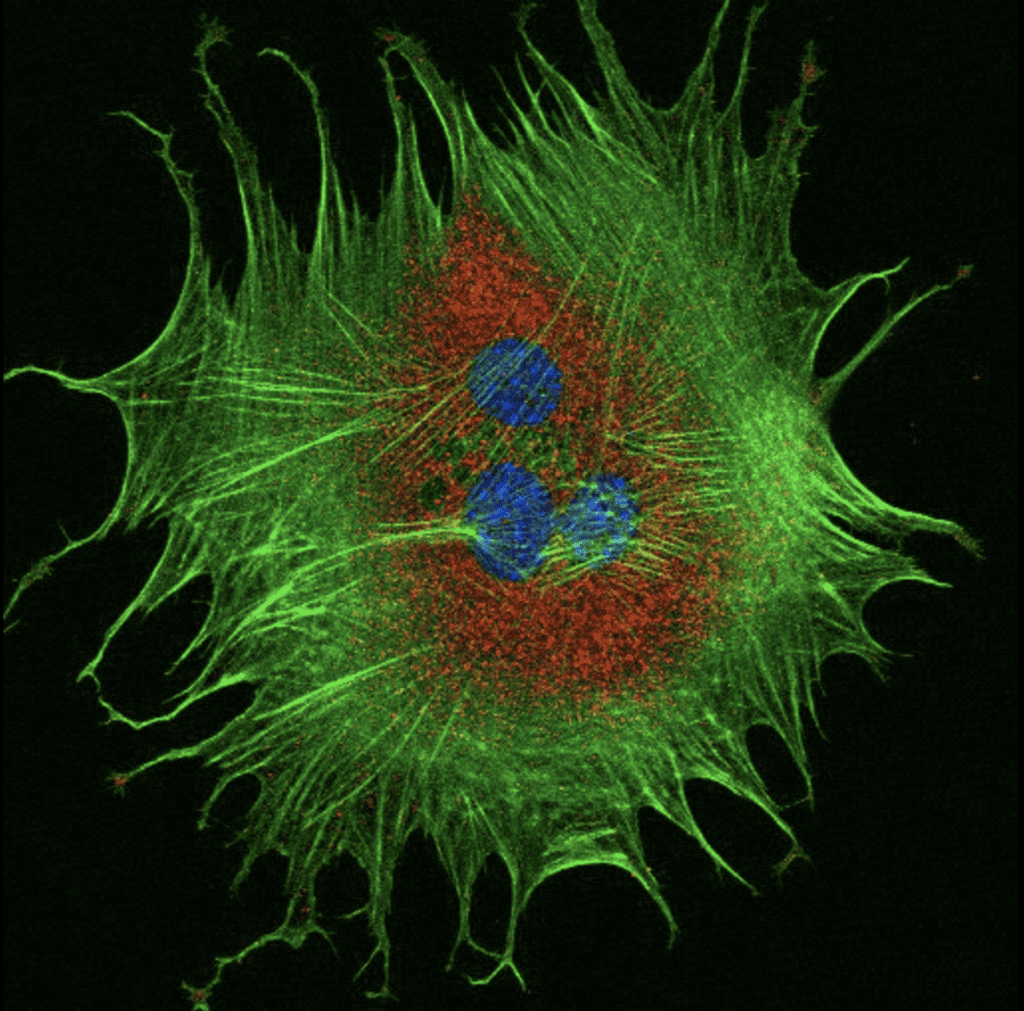
Stretching routine!
– Juan González Valdivieso, University of Glasgow
Myoblasts spread on stiff polyacrylamide hydrogels. The substrate is rigid enough to allow the cells to spread and migrate. The experiment is included in a project to study the cell-biomaterial interaction to develop 3D scaffolds to promote myogenic differentiation in patients with muscle dystrophies. Blue: cell nuclei; Red: vinculin (focal adhesions); Green: actin filaments (cytoskeleton).
For the Love of Bone
– Maria Florez Martin, University College London
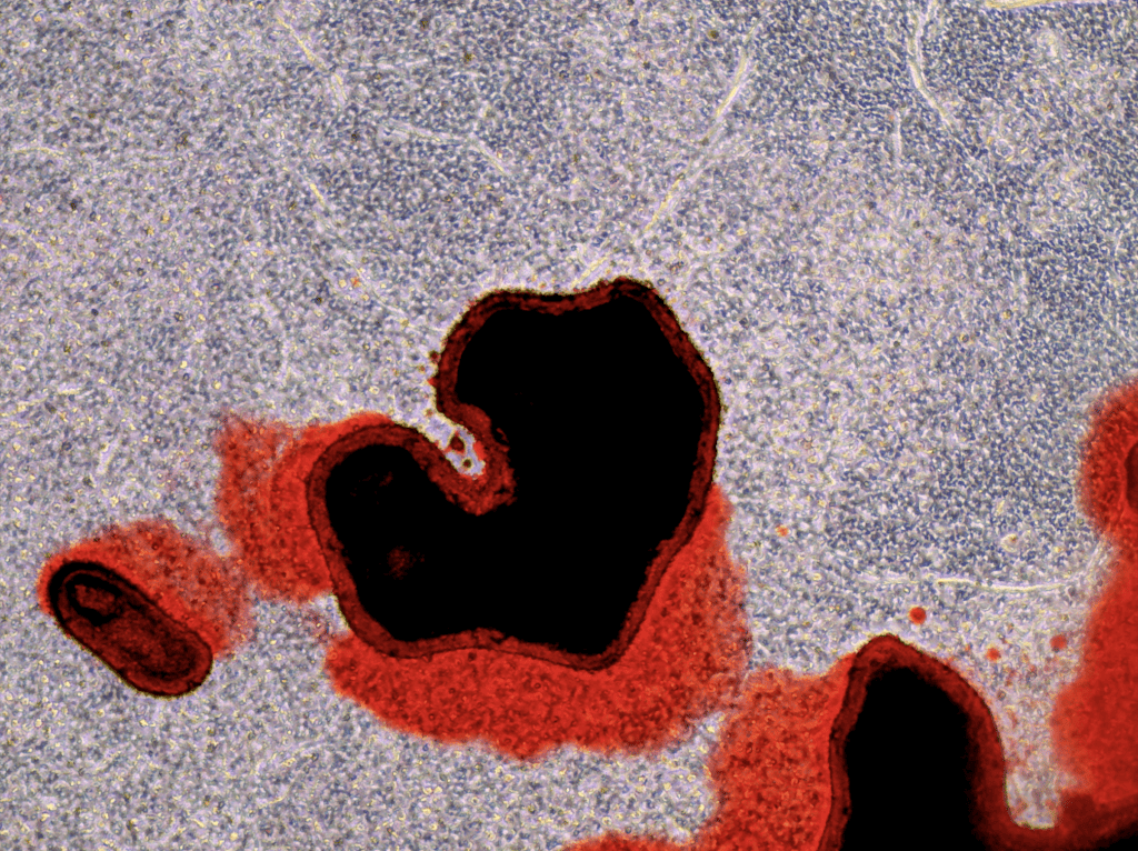
This heart-shaped lab-grown bone, formed in a dense collagen gel with primary rat osteoblasts, is a testament to the love and dedication I have for my work. After 21 days of growth, the bone was stained with Alizarin Red, revealing a beautiful structure highlighting the calcium deposition process, and the intricate beauty of science.
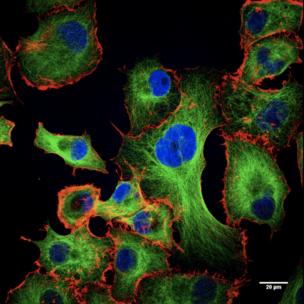
We like to move it!
– Pilar Acedo, University College London
We all travel and cells in our body move too. The cytoskeleton helps cells maintain their shape but also enables cell division and movement. This is key in processes like metastasis. This image shows two components of the cytoskeleton, microtubules (green) and actin filaments (red). I study how different treatments alter proteins of the cytoskeleton of pancreatic cancer cells.
The light of chemistry
– José Luis Marín Rubio, Newcastle University
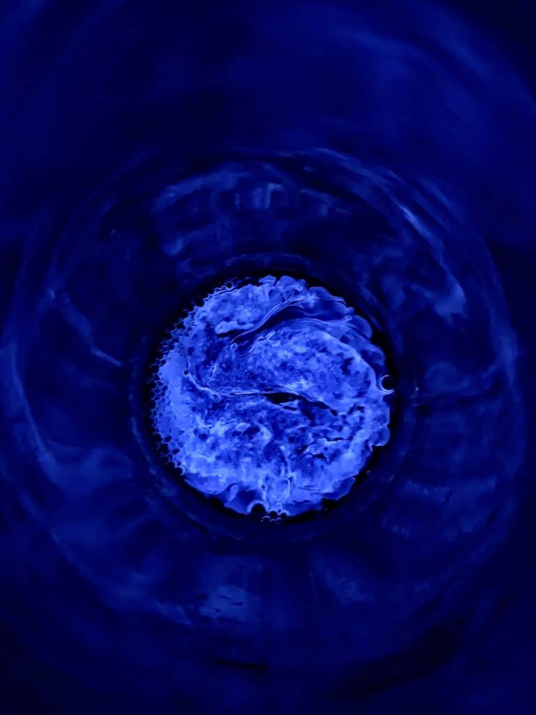
Fluorescence is a widely used tool in the daily life of the laboratory, from fluorescent proteins to chemical reactions. Here the luminol reacts with hydrogen peroxide and becomes dianion and emits flashes of blue light.
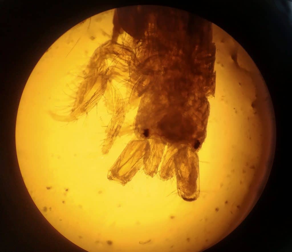
Marine life through the lens
– Loreto Gestoso, Environmental consultant
Amphipod head through a compound microscope. To identify some species of benthic animals, researchers need to use compound microscopes to look for specific parts of the anatomy that identify which species they are.”