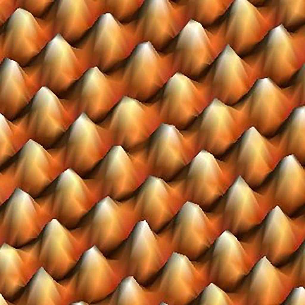
Feel the energy to observe the invisible
Like if were small mountains or Lego pieces. This is a graphite surface where each carbon atom looks like a small mountain, with a lighted peak due to the atom´s localized electrons. Nanotechnology techniques allow scientists to “touch” these electrons and, therefore, to see and manipulate atoms like Lego pieces. A nanoscale regime where everything is different and where the “invisible” revolution of the scientific world is happening.
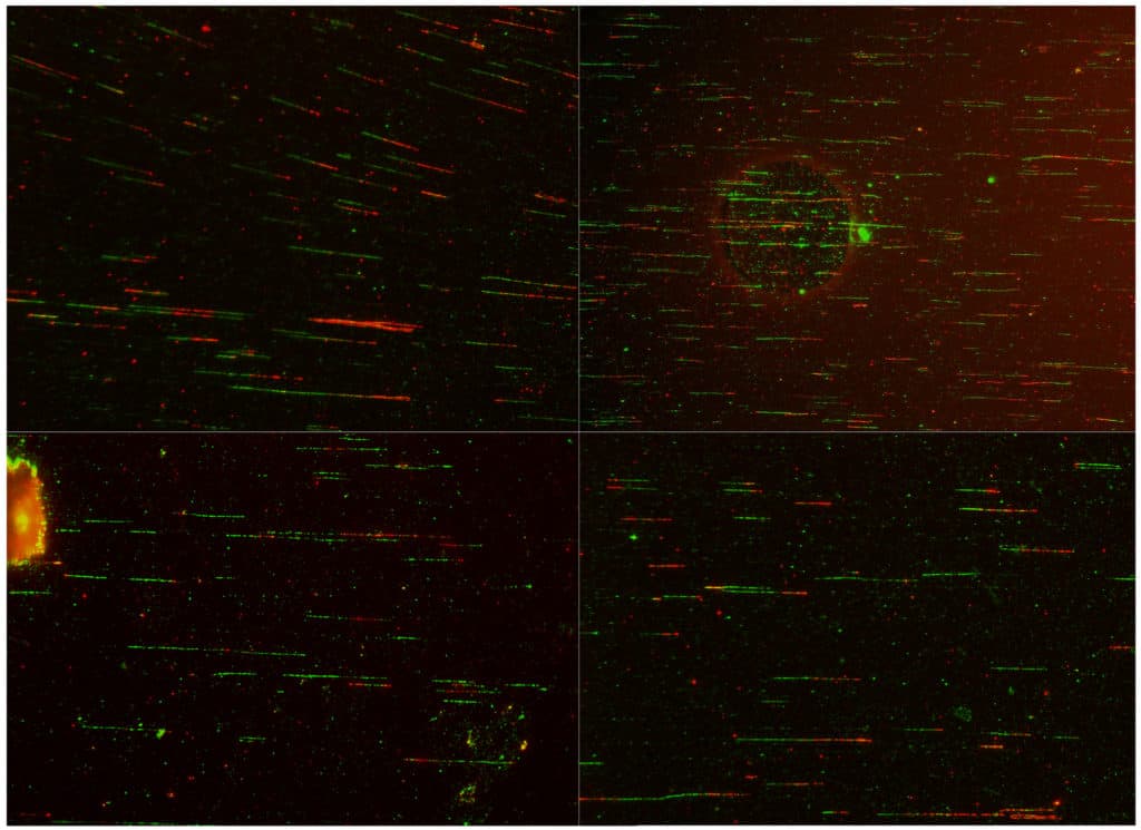
DNA Cosmos- Human DNA captured during replication
Immortalized human cells were labelled with nucleotides (green), treated with cancer drug and labelled again (red). Cells were collected and DNA combing and immunostaining protocols followed. Only replicating DNA is visible. By measuring the strands we can detect replication problems. This special selection of pictures includes artifacts together with the DNA, closer to real science and further resembling the cosmos.
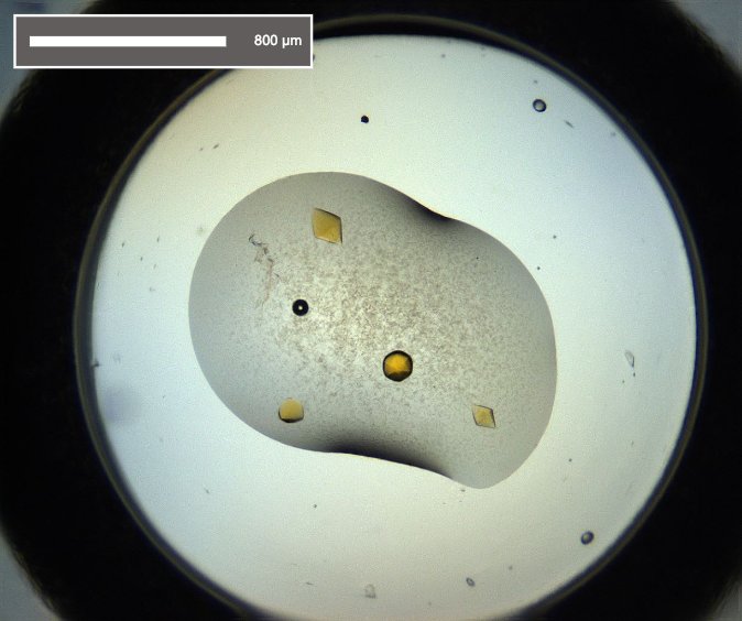
Golden Diamonds
This photo shows protein crystals from Pseudomonas auroginosa reductase. The yellow color comes from the oxidized FAD, a cofactor that is always bound to the protein. The protein molecules organise into the space group P61 resulting in the diamond shape.
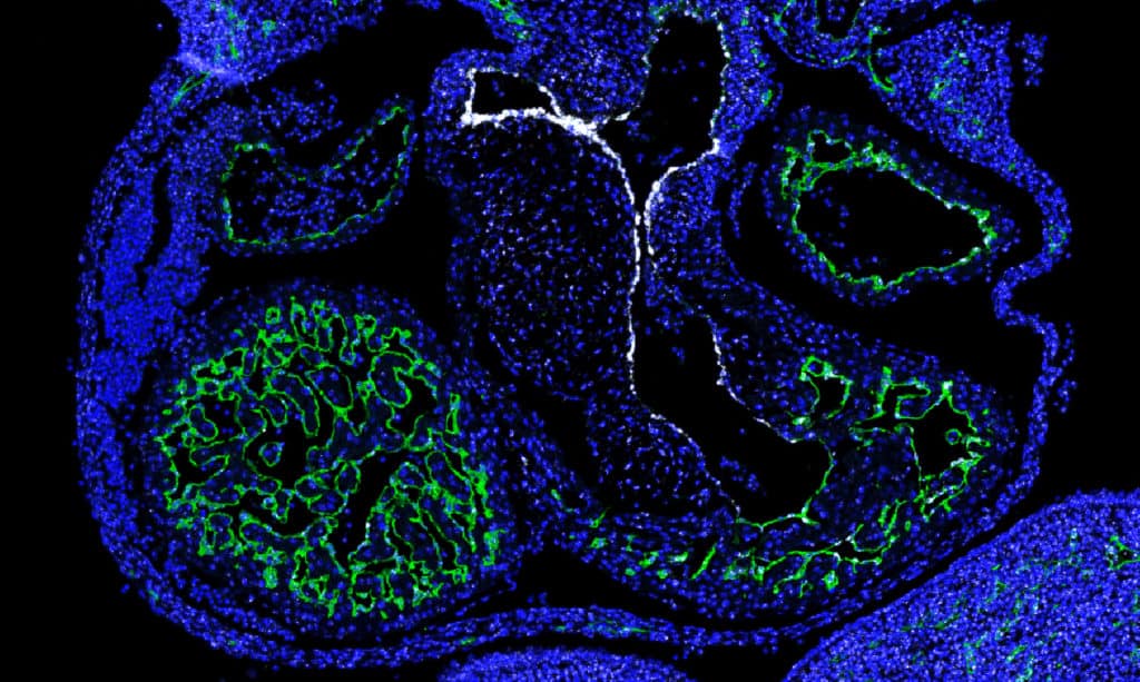
My heart just flows thanks to you
Transcription factors control gene activation/suppression leading the development of different cell types. This image displays an embryonic heart, whose lining is formed by endothelial cells (green). A specific subset of these cells expresses the transcription factor NFATc1 (white). NFATc1 drives these cells to form the aortic valve that will control heart flow from the left ventricle into the aorta.
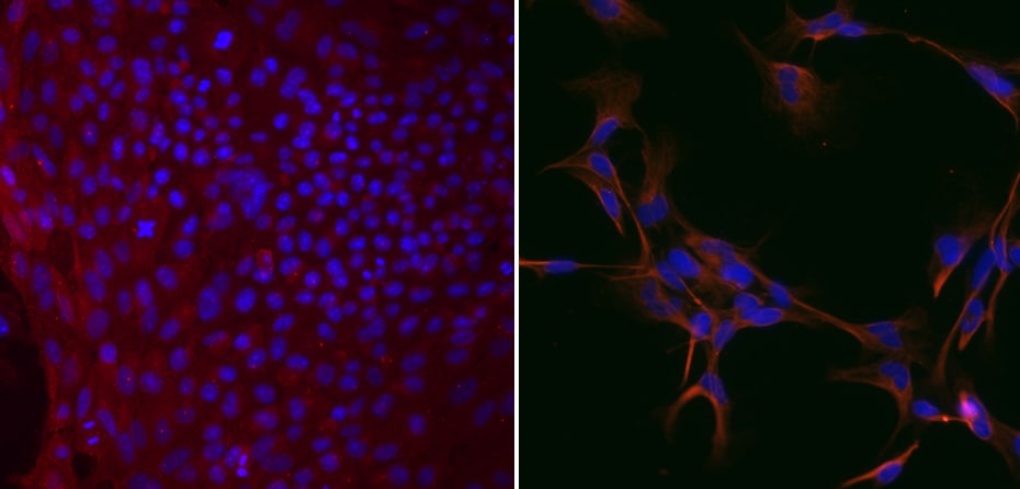
Neural differentiation of induced Pluripotent Stem Cells
Stem cells, specifically induced Pluripotent Stem Cells (iPSCs), were differentiated into Neural Progenitor Cells (NPCs), a stage before neurons. These both images were obtained by immunofluorescence staining of iPSCs (left) and NPCs (right) with stem cell and neuronal markers (both in red), respectively.
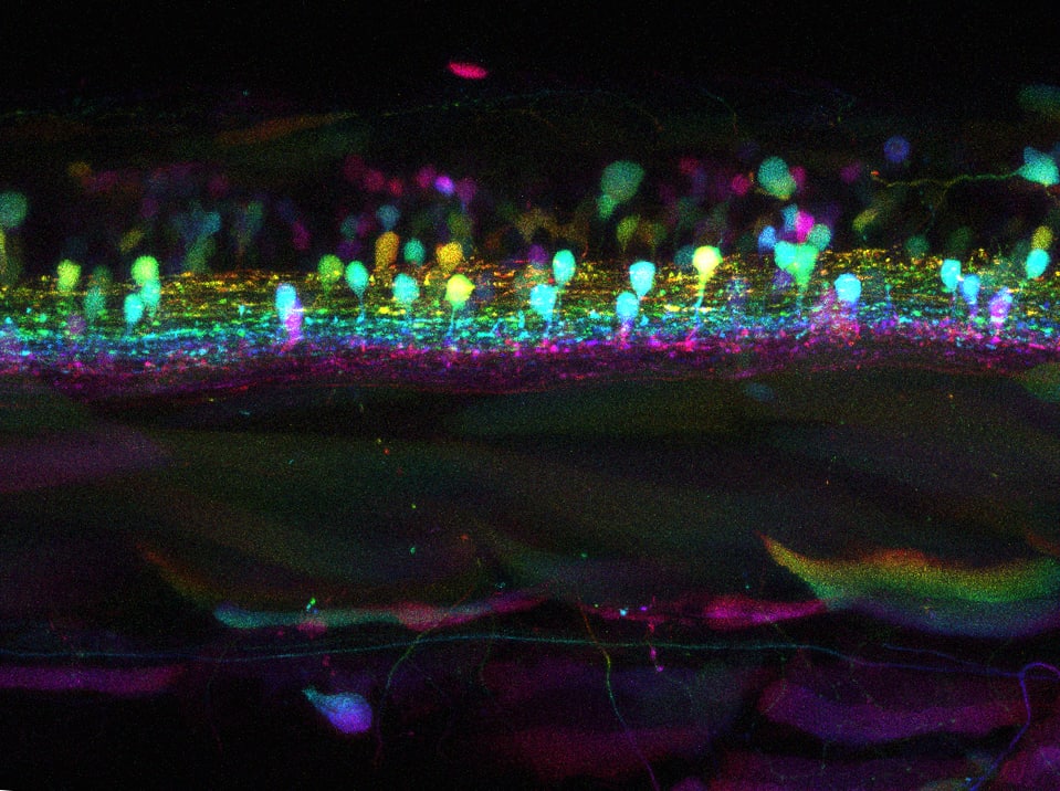
Garbage at the Spinal Cord Disco
Despite the large variety of neurodegenerative diseases, all share a common feature: the accumulation of toxic proteins. This picture shows bright neurons seen at different depths (color-coded) and their axonal-tracks across the spinal cord of a living zebrafish embryo used to study Parkinson’s Disease. Puncta in axons, far from the beauty of colorful dots, correspond to aggregates of toxic alpha-synuclein.
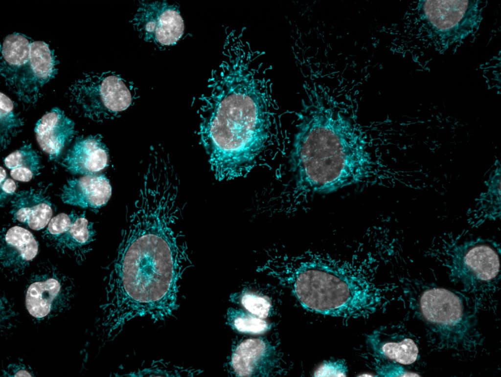
We got the power!
Bile duct cancer cells stained with a fluorescent probe highly specific for the complex mitochondrial networks (in blue). These are key organelles that act as the powerhouse of cells. Labelling these organelles allows for the design of novel therapies that target energy production to inhibit cell division and growth. Nuclei are counterstained in grey.
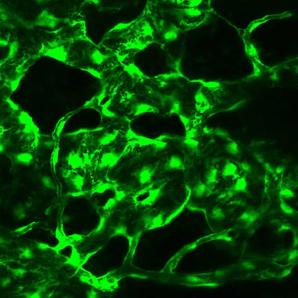
Lost tracks
The image, acquired by intravital microscopy, shows cells lining bone marrow microvasculature (in green) from a mouse. It is dilated, and structurally altered after a cycle of chemotherapy. Chemotherapy is a double-edge sword, killing cancer cells, but also generating several side effects in patients. Protecting and rescuing vasculature improves chemotherapy delivery, decreasing its toxicity and side effects.
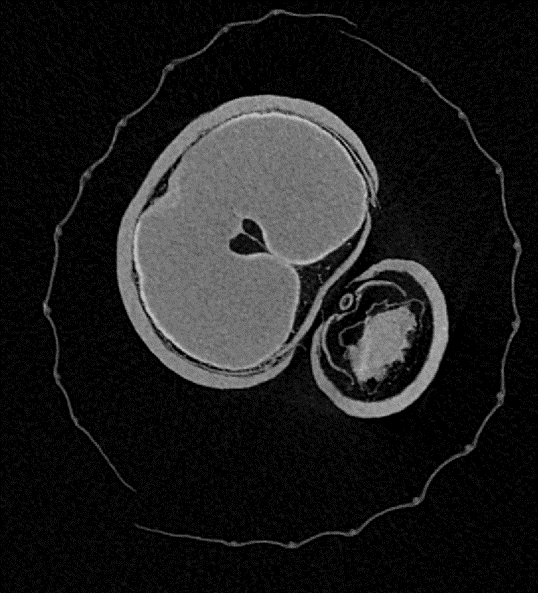
Inside out
Photograph of a section of a winter oat Spikelet through the micro-CT scanning. The section shows the outer layer, the glume, two grains inside, to the left primary and to the right secondary, protected by a leaf like structure, the husk. From Analysis of the genetic and environmental factors affecting grain quality in oats.
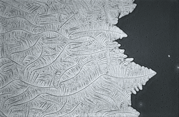
Trees of Salt
We are used to feeling the salt crystals forming on our skin after swimming in the sea, but we are not aware of how this salt is tattooing our skin at the microscale. When water evaporates, salt crystals begin to appear after the formation of small nucleus that quickly starts to ramify, creating intricated branches like the ones in trees.
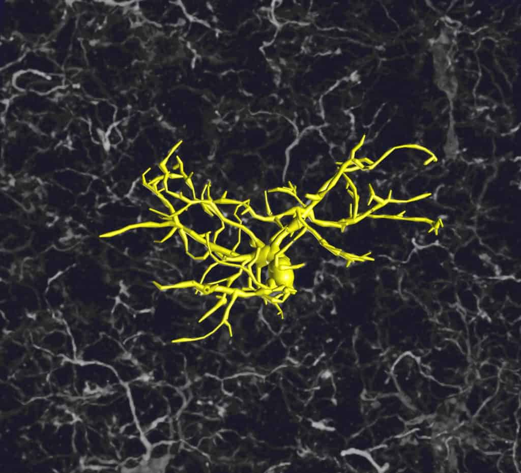
Microglia
Microglia are immune cells which inhabit our brain and constantly scan it with their tiny tentacle-like branches. When they detect a threat (like bacteria or clumps of toxic proteins in dementia), these sentinels rapidly change their shape to respond. Here, one of these cells in a mouse brain has been drawn in 3D to study its structure.
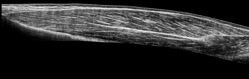
Drawing a muscle
Ultrasound imaging allows to picture tissue inside the human body. In this picture we can see the Vastus Lateralis of a young healthy male (Tissue between the first two white bands). The diagonal white lines are groups of muscle fibres named as muscle fascicles. The length and inclination angle of the fascicles can be used to understand the functionality of the muscle tissue.