In the time it takes you to read this post, 3,500 selfies will be taken around the world.
Each of these selfies will be created by turning the light that arrives to our camera into tiny electrical impulses. These electrical impulses will be translated into the “computer’s language” of ones and zeros and will construct a new digital image ready to be filtered and uploaded to social media. However, these selfies only capture our outside – what would we see if we could take a selfie of our inside? Imagine for a second that we could take a “picture” of a drop of our blood, with a camera really similar to the cameras inside our phones. What could we learn from this little drop of blood?
By mimicking the physical principle used in our phone’s camera, our blood and fluids can be photographed by using microscopically small sensors called ‘ISFETs’. These ISFETs detect the ion concentration in the bodily fluids and create a small electrical current when this concentration changes. Just like cameras use thousands of pixels to achieve a complete image, this “camera of our insides” uses different pixels to detect the concentrations of different ions in our blood: calcium, potassium, sodium, etc…
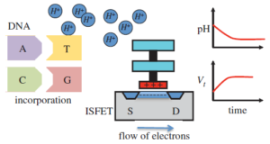
ISFET diagram (From Toumazou C, Tan Sri KT Lim, Georgiou P. 2014. A new era of semiconductor genetics using ion-sensitive field-effect transistors: the gene-sensitive integrated cell. Phil. Trans. R. Soc. A 372: 20130112)
Of the available ions, hydrogen (H+) is the most important one, as it allow us to take “pictures” of our genetic material: our DNA. By taking several ISFET photographs of DNA in our experiment (for the tech-savvy, a DNA amplification), we can see that every time two pieces of the genetic material join together, our ISFET will detect a change in H+ ions. Hence, by recording these ion changes, a “video” of our DNA can be obtained. Unlike with previous technologies, this video can be created in a matter of minutes, very much like how we would record it with our phone.
These “DNA videos” have the potential to change healthcare and the way we diagnose diseases like for example the Zika virus. Imagine for a moment that, after few days on a tropical country during your holidays, you start feeling sick and your body temperature rises. By using these ISFET photographs, you could monitor the H+ ions when mixing a drop of your blood with a set of “templates” of tropical diseases’ DNA. If these photographs show changes in ion concentration, it means that the “templates” matched with a pathogen in your blood, and there is a little bit of disease DNA inside your body. In other words: you would need to see a doctor urgently!
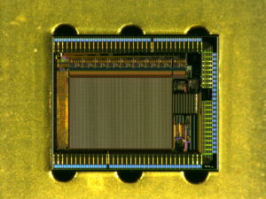
My latest ISFET Chip
These diagnoses are especially important in rural areas in developing countries, where access to diagnostic centres is very limited. In these situations, the possibility of obtaining a fast, reliable and cheap diagnosis in your hometown can make the difference between life and death, potentially saving thousands of lives every year.
Using this new technology, we can now imagine a future in which doctors will take “photographs of our inside” to understand their patients, providing all necessary information to give a precise diagnosis and personalised treatment. A future in which infectious diseases in developing countries could be diagnosed and controlled using a simple and inexpensive DNA test. A future in which “selfies” save lives.
Post by Miguel Cacho Soblechero, PhD Student at Imperial College London. SRUK London Constituency.